The aluminium industry never shies away from telling friendly ears that aluminium is biologically benign. Open ears flap to their message that aluminium is biologically unreactive and so essentially non-toxic. Well this lack of toxicity only applies to humans since they, the aluminium industry, do not deny aluminium’s role in the death of fish and trees brought about through Acid Rain. Well, that is a ‘natural phenomenon’ I have heard them retort when questioned on their blinkered views.
For many years the aluminium industry defended their opinion that aluminium was not even present in the human body at least not in any significant amount. The industry is particularly defensive about aluminium in human brain tissue and has supported various propaganda campaigns involving bought ‘scientists’ claiming all reports on aluminium in human brain tissue were due to issues relating to contamination. We brought that defence to an end and myriad quality publications now leave no doubt on this issue. Aluminium is present in every tissue in the human body, see for example a recent paper on this subject. However, what remains to intrigue us is how does it get there and this is the subject of today’s musing.
Let it be clear, the biologically reactive form of aluminium is its free solvated metal cation, Al3+. You know this from many of my previous posts. What you may not know is that biology abhors any M3+metal cation. The only exception to this is iron, Fe3+ but iron is redox active and biology uses iron as Fe2+and only stores iron as Fe3+. I will come to the significance of this biology in a future musing.
So, there are no metal cation transporters in biology capable of dealing with Al3+. You can read about this here or in my book. This means that unlike all essential charged metal cations there are no easy routes of uptake of Al3+ into cells. So how does aluminium gain access to the human body?
We will examine this through the use of a number of schematics.
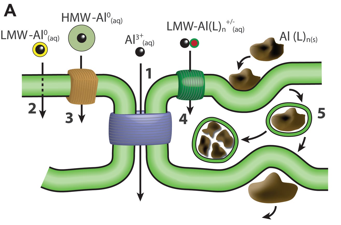
Scheme 1
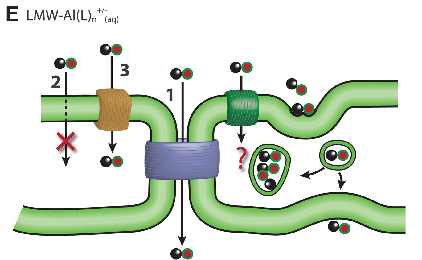
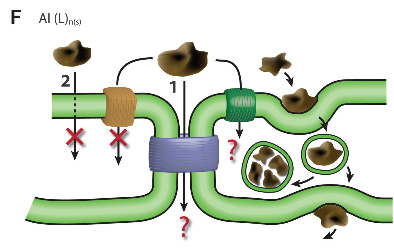
Here we identify five mechanisms whereby aluminium might enter a cell, become intracellular, and/or cross a membrane.
Paracellular route - aluminium squeezes between adjacent cells.
Transcellular route - aluminium diffuses across the cell membrane.
Active transport - aluminium is actively (requires energy) transported across the cell membrane.
Channels - aluminium diffuses through pores or channels in the cell membrane.
Adsorptive or receptor-mediated endocytosis - aluminium is carried across the cell membrane inside a membrane vesicle.
The schematic also identifies five different forms of aluminium, including Al3+, whereby aluminium might cross a cell membrane. We will look at each of these in the following schemes.
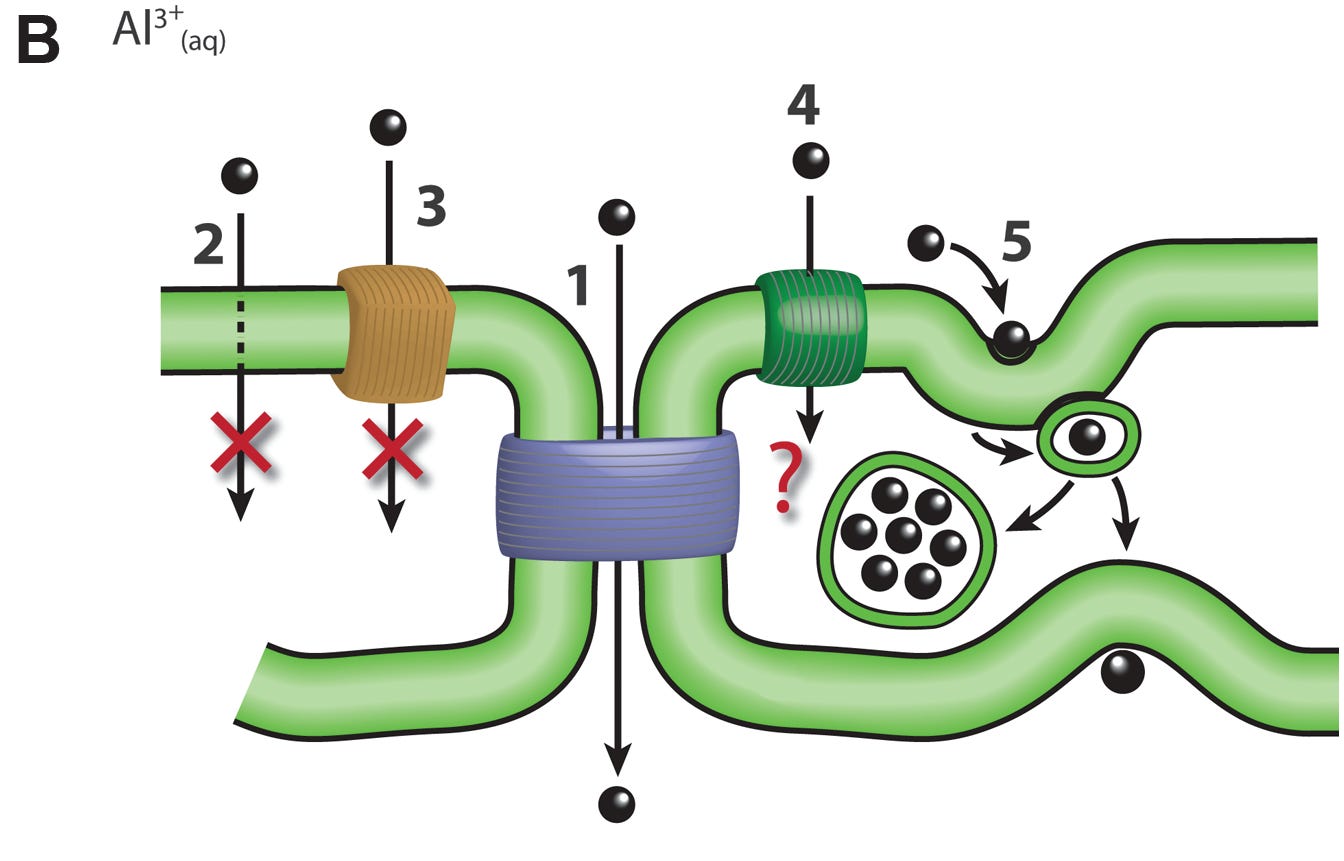
Scheme 2
The schematic suggests that only routes 1 (paracellular) and 5 (endocytosis) might be amenable to the transport of Al3+.
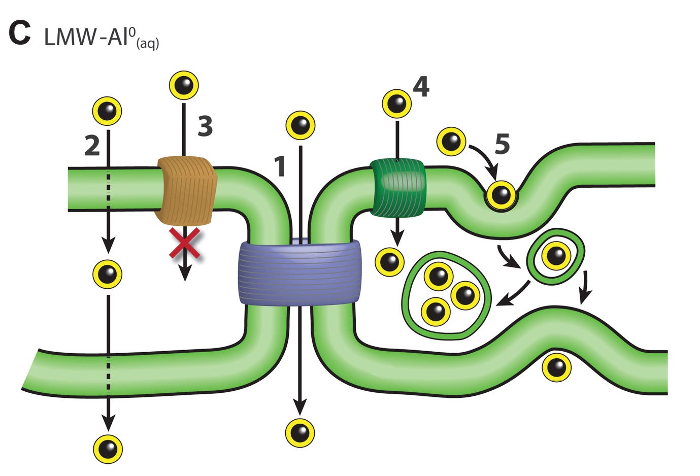
Scheme 3
The schematic suggests that the transport of uncharged low molecular weight soluble complexes of aluminium is only unlikely to occur via an active transport mechanism.
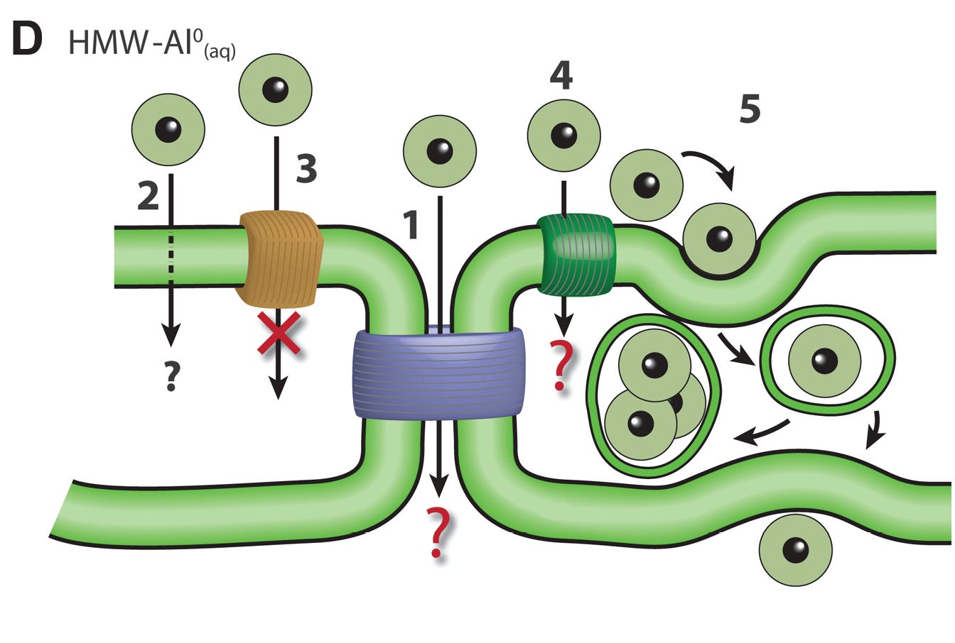
Scheme 4
The schematic suggests that uncharged high molecular weight soluble complexes of aluminium are only likely to cross a cell membrane by a process involving endocytosis.
Scheme 5
The schematic suggests that charged (carrying a positive or negative change) low molecular weight soluble complexes of aluminium may cross a membrane using the paracellular route, a mechanism involving active transport and a mechanism using endocytosis.
Scheme 6
Finally, this schematic suggested that particulate aluminium, for example as is found at the injection site of an aluminium-adjuvanted vaccine, will only pass across a cell membrane by a mechanism involving endocytosis.
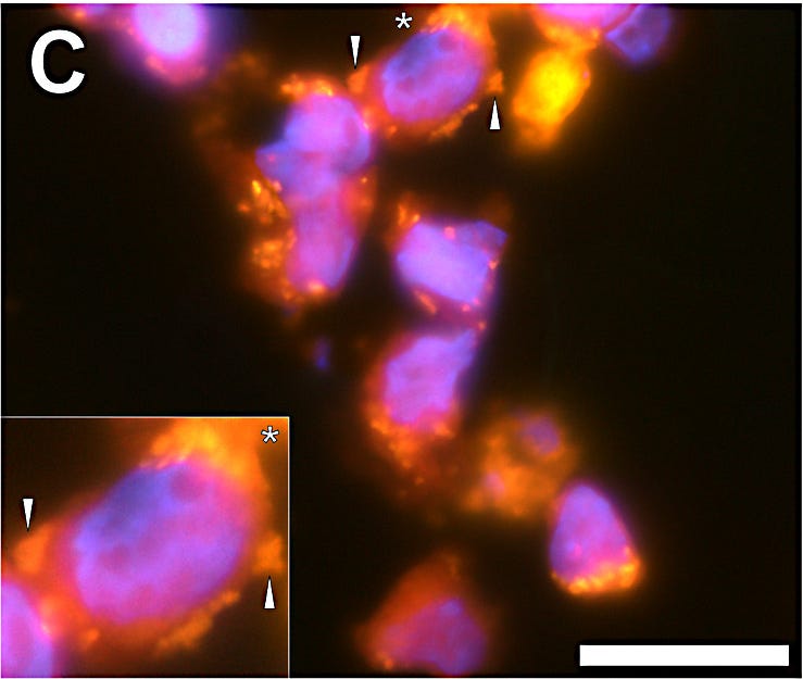
I trust that this series of schematics leaves you in no doubt as to the potential complexity of the transport of aluminium across cell membranes. There remains much to understand though what we know for certain is that aluminium does gain entry to each and every cell in the human body. Once entry is achieved, for example as we have shown in cells present at the injection site of a vaccine, see below, aluminium will wreak havoc.
The image shows THP-1 cells (monocytes) following exposure to aluminium adjuvant. Aluminium, fluorescent orange, is found both inside the cells (intracellular) and associated with the membrane of the cells. Blue fluorescence depicts the cell nucleus.
Dr’s Newsletter is brought to you free of charge, both reading and commenting. However, should you feel that my efforts in bringing to you the story of aluminium are deserving of reward then do feel free to take out a paid subscription. It will be very much appreciated.


The number of bought scientists and doctors is mind boggling. They have sold out their families and friends, surely realising they will not be “saved” for long. Thanks for all the information and schematics that help my brain understand. 👏👏
Aluminum adjuvant = Autism