My rapid removal from academia in August 2021 left our ongoing research into Parkinson’s disease wholly in the lurch.
Since I am no longer in position to publish these data in the usual way I have decided (with the agreement of fellow members of my team) to share them with you on Dr’s Newsletter.
My intention is to simply post the unpublished data. I do not propose to provide in depth analyses of the data. Any readers of my substack with experience in Parkinson’s disease whom would like access to the data may express this interest through commenting on the relevant post.
The magnificent microscopy to follow was undertaken by Dr Matt Mold, my long time collaborator who also lost his job when I left academia in 2021.
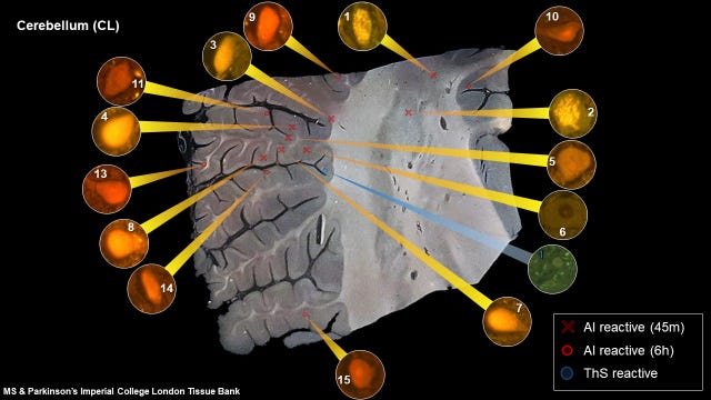
The first image is a section of the cerebellum of a 74 year old man with Parkinson’s disease.
The structure and function of the cerebellum is compromised in Parkinson’s disease with particular reference to tremor.
Purkinje cells are specific targets in the cerebellum in Parkinson’s disease.
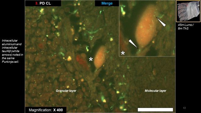
This image displays orange fluorescence depicting the presence of aluminium and green fluorescence identifying amyloid. The large Purkinje cell in the centre of the image between the granular and molecular layers is showing significant positive fluorescence for aluminium. The Purkinje cell is also showing positive fluorescence for amyloid, one possible form of amyloid known to deposit in Purkinje cells is alpha synuclein. Other green amyloid-positive fluorescence in the region between the granular and molecular layers might be amyloid-beta or tau.
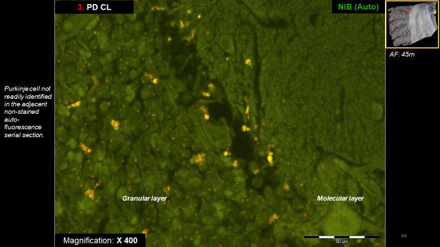
Just for those Doubting Thomas’s (and you should always question the science) the next image shows the autofluorescence or control for the same section. While lipofuscin continues to show strong yellow fluorescence neither the orange fluorescence depicting aluminium nor the green fluorescence showing amyloid is visible in the control.
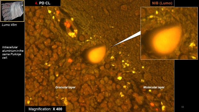
We need to ask how the presence of a significant burden of aluminium affects the structure and function of Purkinje cells, such as shown below.
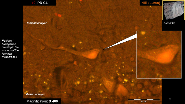
In the following exemplary image of a Purkinje cell and its dendritic root aluminium-positive fluorescence identifies the nucleus of the Purkinje cell.
Research comparing the structure and function of the cerebellum in Parkinson’s disease and in control brains consistently shows fewer numbers of Purkinje cells in Parkinson’s disease. Such an observation could be explained by the presence of cytotoxic aluminium.
I hope that you appreciate me posting unpublished original research on aluminium and Parkinson’s disease. You are the first to see these images and I am making them available to all subscribers. Should you feel that the information I provide is worthy of a paid subscription then know that such is very much appreciated.


My hope is that a new administration in the US, with RFK in HHS, will mean funding for you to continue your research.
It’s Astonishing that such brilliantly advanced research & findings are targetted by Academia to protect the interest of The Pharmaceutical Indistrial Complex at the expence of Folks suffering with Parkinson’s Disease. This is what Academia has sunk to.
Question Dr Exley : Will folks in thier early 60's who have been diagnosed with Parkinsons ( & are on Pharma Pills ) Still benefit from Drkining Silica Rich Water , Like Fiji ? is it ever Too late?
& lastly How may I make a humble donation please ? Sunstack only facilitates Monthly Sunscriptions & yet , many might want to also, make a donation?