It Started With a Fish
Lessons learned from fish gills
Those hardy few who have read my book will know that I began my research on aluminium studying fish. While I was, and remain, a fisherman I never really considered myself a fish biologist. I was hooked on aluminium and fish, being the victims of Acid Rain, were the obvious animal model. I set myself the challenge of understanding acute aluminium toxicity in fish, why aluminium killed fish within relatively short, hours to days, periods of exposure. At the time the convenient consensus was that aluminium was simply a physical toxicant, coating the surface of the fish gill and thus preventing its function.
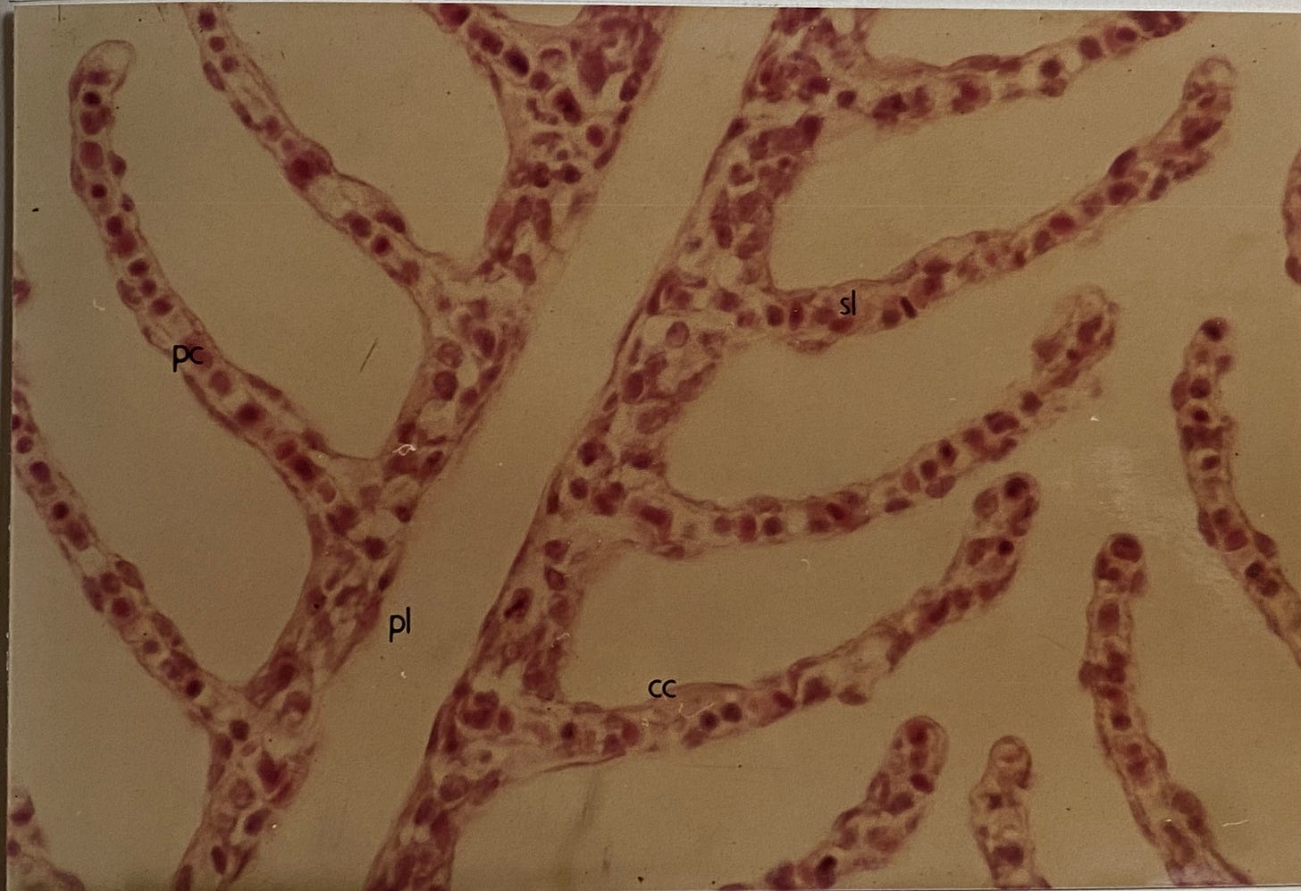
The fish gill is an epithelium with a number of critical functions. It is a respiratory surface allowing for the exchange of oxygen and carbon dioxide. It is an ionoregulatory surface facilitating the movement of ions, cations and anions, into and out of the blood and it is osmoregulatory, controlling the movement of water. At this point I deemed it useful to show you an image of a fish gill. However, I quickly realised that I have not published any images of normal fish gill. Delving deeper I wondered if a digital copy of my PhD existed. This has multiple images of fish gills. I found out that the University of Stirling had produced a digital copy. Alas, a digital copy where all photographs were blacked out. Note that in 1989 photographs were taken on a camera with normal film, developed and then stuck with glue into the thesis. How times have changed. However, I found an excellent photograph of a fish gill in my undergraduate thesis, written in 1984, and I have managed to reproduce this for you below.
Some explanation is required. The photograph is a light microscopy image of the fine structure of a fish gill. It is composed of primary lamellae (pl) and secondary lamellae (sl) creating a vast surface area over which water flows and gases and ions can be exchanged. This image also shows chloride cells (cc) and these allow anadromous fish such as salmon to pass between fresh and seawater and vice versa.
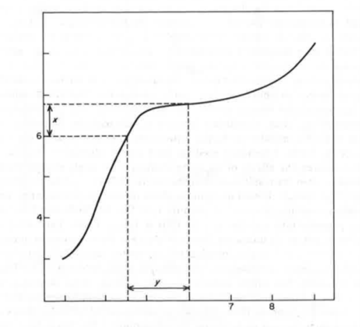
My task was to understand how aluminium disrupted the function of the exceptionally specialised fish gill. My epiphany was in being the first to appreciate something that I called the gill boundary layer (gbl). This is the layer of water closest to the epithelial cells that constitute the gill surface. It is the layer that dictates the chemistry at the gill surface and therefore the chemistry of aluminium at the gill surface. This idea is depicted in the image shown below.
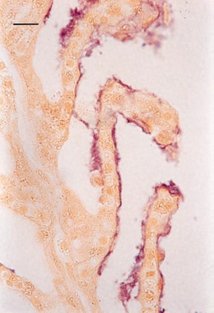
The figure is taken from the paper in the previous link. It is incomplete, it does not look like this in the paper (please ask if you would like a copy) but pdfs of papers published in the early 90s are not so easy to edit! The x-axis (horizontal) depicts the pH of the bulk water, the water the fish is swimming in. The y-axis (vertical) depicts the pH of the gill boundary layer (gbl). The figure shows that when the pH of the bulk water is between 4.5 and 6.0 (labelled ‘y’ in the figure) the pH of the gbl, the layer of water closest to the gill surface is circumneutral, between pH 6.0 and 7.0 (labelled ‘x’ in the figure). The reason for this is a combination of factors that I discuss in the paper but primarily the pH of the gbl is determined by the dissolution at the gill surface of excretory products, carbon dioxide and ammonia, and their competing equilibria. The importance of this equilibrium between the acidic carbon dioxide and the basic ammonia is further reinforced by a stream of mucus across the gill surface. An example of this mucus stream, identified by a blue stain, is shown in the image below.
The figure shows a primary lamella with secondary lamellae all seemingly coated with a steady, dynamic stream of mucus.
The fact that the gill boundary layer remains at circumneutral pH even when the bulk water pH falls below pH 5.0 explains why mild acidity per se is not toxic in fish. However, bulk water at pH 5.0 that also includes 200ppb total aluminium is acutely toxic within 24-48h. The exact mechanism of acute aluminium toxicity under these conditions is explained in detail in the aforementioned paper but in brief the actions of aluminium, acting through Al3+, disrupts the integrity of the gbl and binds at functional groups at the gill surface. These actions of aluminium are shown in the image below, aluminium is stained purple.
Aluminium is shown bound at the surface of the gill and also associated with the gill mucus. I will re-visit the subject of aluminium and mucus in a future substack post. The disruption of the structure of the gill lamellae is already evident in this image.
The fish gill is an epithelium and is similar in form and function to many other epithelia including those in humans, for example, in the gastrointestinal tract and the lung. Studying how aluminium interacted with the fish gill epithelium taught me a great deal about how aluminium interacts in biology in general. It continues to inform my thinking and my learning and it all started with a fish.


I remember in one of your excellent presentations about this mechanism in fish, a pediatrician in the audience wondered if this mechanism could also be affecting children and elderly who receive the vast majority of Al containing vaccines and whether it is related to the asthma, COPD and other breathing conditions affecting highly vaccinated humans!
Fascinating. Always love to understand the mechanisms (engineer talking). It provides so much useful knowledge. I did wonder whether that boundary layer you found was (what has now been trumpeted as a major factor) by the likes of Prof Gerry Pollack as EZ (Exclusion Zone) or structured water. They sure look very similar, from your description and fulfil the same functional requirement of being part of trans membrane transport.